Macular Degeneration
Macular Degeneration, also known as Age-related Macular Degeneration (AMD), is a condition that can cause vision loss in the area of the eye responsible for sharp, central vision. AMD is a progressive disease that primarily begins later in life (usually after the age of 55) and impedes vision, sometimes to the point of legal blindness. Currently, AMD affects 15 million people in the United States, which is more than Cataracts and Glaucoma combined.This article will provide the tools, information, and knowledge needed to detect AMD early, prevent progression, and understand key risk factors. In addition, you’ll learn about how assistive device technology can help those living with AMD.
Overview
What is Age-Related Macular Degeneration?
Macular Degeneration, also known as Age-related Macular Degeneration (AMD), is a common condition that causes central vision loss and “fuzziness” of vision.
AMD is caused by the deterioration of the macula located within the center of the retina at the back of the eye. The macula is responsible for central vision, which is what you see in front of you when looking straight ahead. There are two types of AMD: wet Macular Degeneration (wet AMD) and dry Macular Degeneration (dry AMD). Both wet and dry Macular Degeneration can cause substantial vision loss.
Age-related Macular Degeneration as a type of progressive vision loss can make everyday activities very difficult, such as reading, driving, and recognizing faces. However, there are ways to prevent the development of AMD and slow the progression of this disease.
The ICD code for AMD is H35.30.
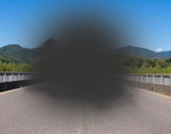
An example of what Macular Degeneration looks like.
How Does AMD Affect the Eye and Central Vision?
Within the eye, there are three main layers which lay flat against one another to form the eyeball:
- Sclera – the “white” part of the eye
- Choroid – the middle layer of the wall of the eye, between the sclera and the retina
- Retina – paper-thin, light-sensitive tissue lining the inside of the eye
Light from the outside world is used to create visual information for the eyes and create vision. After light enters the eye through the cornea, the retina absorbs the light and changes the visual information into electrical impulses. The optic nerve at the back of the eye then carries these impulses to the brain, which decodes the impulses into a visual image. Macular Degeneration creates vision loss by affecting the retina’s ability to absorb light effectively. Macular Degeneration specifically affects the macula, which sits at the center of the retina. The macula contains the highest concentration of cones, which are photoreceptor cells responsible for providing sharp, detailed central vision. AMD causes these cones to degenerate in a way that prevents them from absorbing light correctly.
How Common Is Age-related Macular Degeneration (AMD)?
Age-related Macular Degeneration is highly prevalent worldwide and affects most people at the age of 55 years or older. In Canada alone, AMD affects approximately 1.4 million people. About 15 million people experience AMD in America and AMD accounts for 8.7% of blindness worldwide, making it the leading cause of vision loss—more than Cataracts and Glaucoma combined.
Types of AMD
Types of Macular Degeneration
There are two types of AMD: dry Age-related Macular Degeneration and wet Age-related Macular Degeneration.
Dry Age-related Macular Degeneration
Dry AMD occurs when parts of the macula become thin and tiny clumps of protein or fatty deposits, known as drusen, collect in the macula. Dry AMD is considered an early stage of AMD and accounts for approximately 80-90% of all people with Macular Degeneration.
Fortunately, dry AMD typically does not progress further than pigment discoloration and the presence of drusen, and if it does progress, it is usually very gradual. It is possible for dry AMD to progress to wet AMD.
Wet Age-related Macular Degeneration
In cases of wet AMD, the retina becomes affected by abnormal blood vessels growth. These blood vessels leak blood and other bodily fluids that can cause damage to the macula via scarring.
Wet AMD is less common, as it occurs in only about 10-20% of individuals with AMD. However, wet AMD is significantly more serious than dry AMD because it is much more likely to cause severe vision loss. In fact about 90% of all cases of severe vision loss related to AMD are caused by wet AMD.
Vision loss progresses faster in those with wet AMD than it does for those with dry AMD. Wet AMD can also be called neovascular AMD or exudative AMD because it involves the exudation (leakage) of fluid and blood from blood vessels.
Signs and Symptoms
The early signs and symptoms of dry AMD include:
- Increased difficulty with night vision
- Experiencing low vision or blurred vision
- Printed texts appearing blurry
- Visual distortions, such as straight lines appearing wavy
- Reduced central vision in one or both eyes
- Reduced sensitivity to colors
- Unable to recognize faces
Signs and symptoms of wet AMD’s symptoms include:
- Decreased color brightness
- Quick progression of symptoms
- Gray, blurry or dark spots in your central vision
- Visual distortions
- Hazy vision
Symptoms of Age-related Macular Degeneration are usually only noticed once both eyes are affected.
Macular Degeneration is a Progressive Condition
Dry AMD is a progressive disease, which means that symptoms of AMD will get worse over time. However, the disease can advance so slowly that vision loss is not significant for several years although progression can be quicker in more severe cases (most commonly with wet AMD).
Dry AMD does not affect vision in the early stages. However, as the condition progresses, it causes an increasingly blurred area near the center of vision. This blurred area will grow larger over time, and may even create blank spots in the center of the visual field. A common symptom of progression is that objects do not appear as brightly as they once did. Central vision may ultimately be lost; although peripheral vision typically remains. In some cases, people with advanced AMD are considered legally blind.
Age-related Macular Degeneration does not always happen to both eyes simultaneously. It is possible to have AMD in only one eye, or to have different stages of AMD or types of AMD in each eye.
It is important to remember that not everyone with AMD will develop advanced AMD and that there are ways to slow the progression once diagnosed.
Stages
There are three stages of dry Age-Related Macular Degeneration. The first stage is associated with very few symptoms, the middle stage includes some or all symptoms, and the last stage denotes a total loss of central vision. Eye doctors do not classify wet AMD within stages, as wet AMD is always considered to be an advanced form.
- 1
Early Age-related Macular Degeneration
During the early stages of AMD, drusen and/or fatty-protein deposits are present under the retina, but no pigment changes are seen.
Most people do not notice any vision loss at the early stage of AMD; however, an eye doctor or an ophthalmologist can still detect AMD at this stage. As such, regular eye exams are highly encouraged to facilitate early detection.
- 2
Intermediate Age-related Macular Degeneration
In the intermediate stage of AMD, larger amounts of drusen are present under the retina, along with pigment changes. These pigment changes are known as “retinal pigment epithelium (RPE) disturbances”, and can cause central blind spots or overall blurred vision to begin to form.
Not all people with intermediate AMD will notice symptoms, but some do begin to experience difficulty with reading, writing, or other detailed work.
- 3
Late Age-related Macular Degeneration
At the late stage of dry or wet Age-Related Macular Degeneration, central vision loss has occurred. This may include blurry vision, or an inability to view anything within the central field of vision. In most cases, peripheral vision remains intact.
Many people do not realize that they have AMD until the damage to the macula has progressed enough to blur their vision. Given this, it is extremely important to have regular visits with an eye doctor for early prevention and detection.
Causes
AMD can be caused by both genetic and environmental factors, including age, race, eye color, smoking history, family history, and obesity.
Age
Age is the most common factor associated with Age-Related Macular Degeneration. Most people begin to see signs of AMD around age 55, but it can technically present at any age. Approximately 2% of people in their 50s have AMD, but this number increases dramatically to about 30% by the time people are 75 or older.
Race
Race is a known risk factor for developing AMD, with white (European descent) people being in the highest risk group. About one-third of people of white descent have the gene that is linked to AMD.
People who are Chinese and/or Hispanic/Latino are also at increased risk of developing AMD. People of African descent, however, have the lowest risk.
Light-Colored Eyes
People who have lighter eye colors, such as blue or green, have a greater chance of developing AMD. This may be because lighter eyes have less pigment and therefore are less effective at deflecting ultraviolet rays from the sun, making light-coloured eyes more sensitive to light.
Smoking
People who smoke are at up to four times the risk of developing AMD compared to non-smokers. This is because smoking narrows blood vessels and subsequently, reduces the supply of blood and oxygen to the eyes.
Family History
It is estimated that 15 to 20 percent of people with AMD have at least one parent, sibling, or another first-degree relative who also has the disease.
Scientists have identified nearly 20 genes that influence the development of AMD. Some of the identified genes influence the immune system, which protects the body from bacteria and viruses, while others trigger inflammation and remove debris, such as drusen, from cells and tissues.
Obesity
Obesity is known to increase risk of early or intermediate dry AMD, both of which can progress to late AMD or wet AMD. Despite this understanding, the precise link between obesity and the progression of AMD remains unknown. It is hypothesized that cardiovascular disease and sedentary behavior may play a role, but this is yet to be confirmed.
Prevention
Fortunately, there are actions that can be taken to reduce the risk of developing AMD, or slow down the progression of the disease. These include:
- Smoking cessation—Stopping smoking (or never starting) can prevent the development and/or progression of AMD. Current smokers have 2-3 times the risk of having AMD than non-smokers.
- Healthy diet— A healthy diet can greatly help people with dry AMD to prevent the progression of AMD. The most important foods to consume are fruit, fish, and dark, leafy greens that are high in omega-3 fatty acids and antioxidant vitamins.
- Reduce refined carbohydrates—Reduce your intake of refined carbohydrates, such as white bread. Research shows an association between diets containing a high amount of refined carbohydrates and the onset and progression of macular degeneration.
- Regular exercise—An active lifestyle can help to boost the levels of antioxidants in the eyes. Some research suggests that people who engage in regular physical activity are 70% less likely to develop wet AMD.
- Routine eye exams—Booking a yearly eye exam is central to the early detections of dry and wet AMD. If detected, you can immediately begin treatment efforts to stem the progression of the disease.